Behind the Scenes of AI-Powered MRI
A deep dive into how MRI and AI power informed decisions and the future of in-ovo sexing.
Oct 22, 2025

Orbem’s Genus Focus for in-ovo sexing has revolutionized the poultry industry, enabling our customers to enhance their operations in terms of efficiency, sustainability, and animal welfare. The Genus Focus scans and sorts eggs on days 11 and 12 of incubation, accurately and non-invasively classifying each egg as male, female, or unviable. But how does this state-of-the-art technology achieve such accurate sexing analysis across various breeds or incubation ages, and what makes it safe to use? In this post, we take a look at Magnetic Resonance Imaging (MRI) and Artificial Intelligence (AI). When combined, these two key technologies allow us to see what is hidden inside and make informed decisions.
See What Is Hidden: Magnetic Resonance Imaging
1. Magnetic
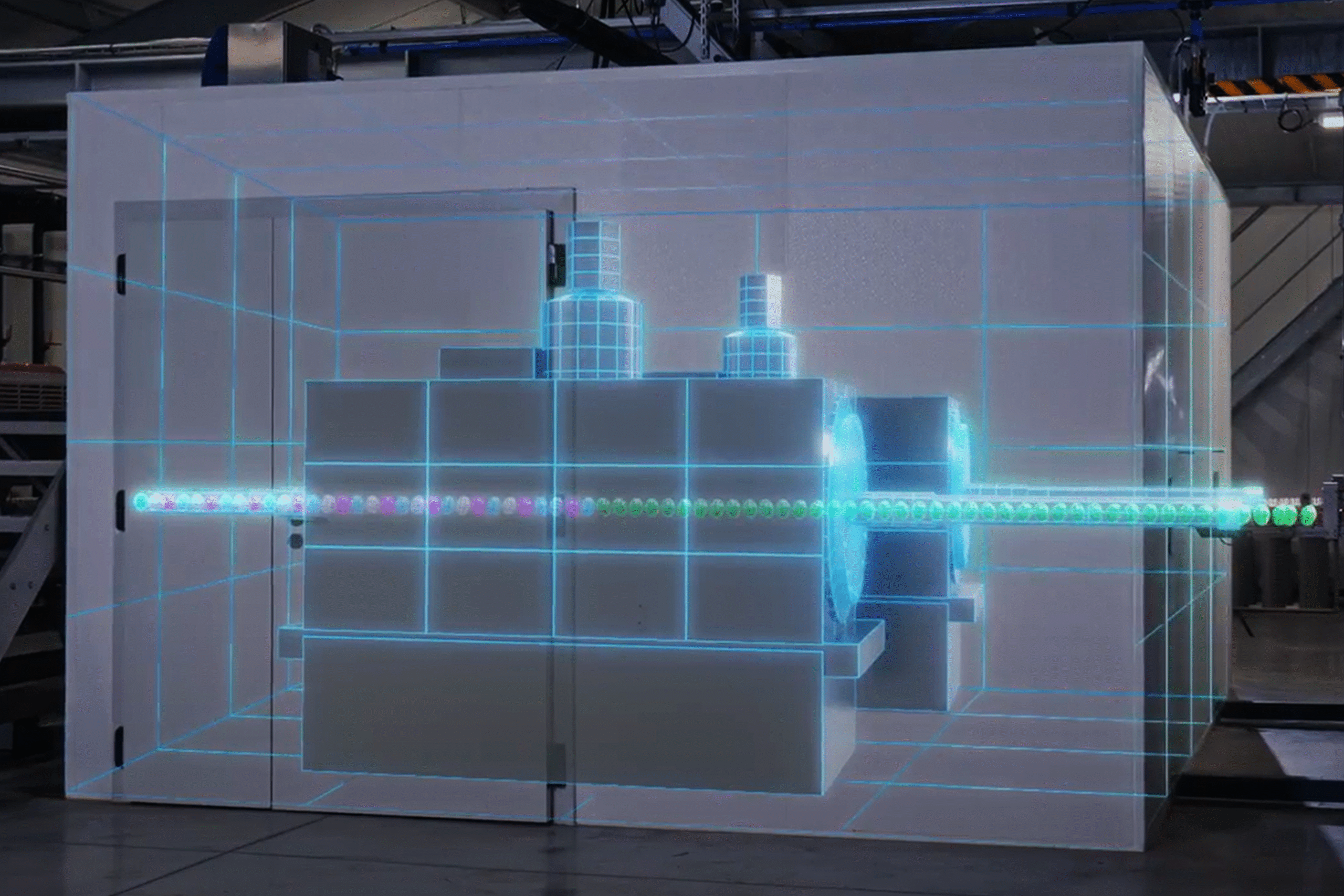
MRI is a remarkably powerful and safe* technique that allows visualization of different tissues and internal structures in human bodies and other objects. MRI scanners generate a harmless magnetic field which interacts with the water molecules of a scanned object. To be more precise, it interacts with hydrogen protons, contained in water (and other molecules such as fat). Given that human bodies are composed of approximately 60% water, they contain an abundance of these hydrogen protons. The same is true for eggs and many other food objects, such as fruit.
* Care should be taken that no metal parts enter the MRI scanner (think of pacemakers or metal-based implants).
2. Resonance
An MRI scanner applies changes to the magnetic field, and hydrogen protons respond to those changes in a way that is called magnetic resonance. Since various body parts (organs, tissues) contain different amounts of water, their hydrogen protons respond to the magnetic field differently, based on the properties of those specific tissues and organs. Essentially, this means that different tissues will appear differently in an MRI image through varying contrast and brightness. For example, bones will have a different contrast than muscles, and similarly, grey matter will be distinguishable from white matter of the brain. Using the same principle, we can see egg white and egg yolk in different contrasts.

3. Imaging
MRI scanners are equipped with sensors which evaluate the resonance of hydrogen protons from different parts of the body and combine this information into a complete 3D image. What does a 3D image look like? Imagine that you make a cut through an apple: you would see a slice (a 2D image) of this particular section of the apple. Now imagine you make another cut – another slice – and then another one. As you cut through the apple, every slice shows you what a different part of the apple looks like.
If you combine those slices together, you get a 3D image. It is an extremely powerful representation that lets you learn about the whole object. Once you have a 3D image, you can even re-slice it at a different angle and inspect shapes and volumes of all internal structures of an object.


Why MRI?
MRI is not the only technology that allows us to see inside objects; however, it offers unique advantages. Unlike X-rays or Computed Tomography (CT), MRI is a non-invasive technology that does not utilize any harmful ionizing radiation. It allows us to see what is hidden in a safe way for products, humans, and the environment. Moreover, MRI works very well in visualizing soft tissues (think muscles and internal organs, or fruit flesh, or egg yolk/white) in contrast to X-ray or CT.MRI excels at producing high-resolution 3D images, revealing anatomical structures, shapes, and full volumes. This capability surpasses ultrasound, which offers lower resolution, and hyperspectral imaging techniques, which provide information only at shallow depths without high-resolution structural detail.
While MRI’s precision and safety have cemented its role in healthcare, its broader application has been limited by high costs, operational complexity, and slow processing times. Orbem is addressing these challenges by industrializing MRI, making it more affordable, user-friendly, and significantly faster. To learn more about how we achieved this and the obstacles we overcame, you can read our amazing colleague Ben Knowles’s blog post on the industrialization of MRI.
MRI allows us to see inside an object and create its high-resolution 3D image. Now let’s see how we use AI to make informed decisions based on those images.
Informed Decisions: Artificial Intelligence
Demystifying AI, ML, DL
The term “Artificial Intelligence” (AI) has become incredibly popular over the last decade.
But what does AI mean exactly?AI is the idea of having a machine or program that can think and act in ways that mimic human intelligence. Imagine a fully automated, “smart” hatchery or sorting robot that can make all its own decisions – for instance, which egg to incubate and which not – without any human intervention.
In the past, programmers had to provide explicit, step-by-step instructions for a machine to make decisions. This often meant painstakingly considering all possible scenarios and exceptions. Sometimes, that task becomes impossible, especially in cases where decisions are to be made based on complex data such as images.
This changed with the advent of Machine Learning (ML), a key method for creating AI systems. In ML, you collect vast amounts of data related to your system – which egg is female or male, humidity, or temperature levels at the hatchery, egg images – and let the system find the patterns in the data on its own. For example, an ML system can analyze past data and learn to predict which eggs are most likely to contain a female or male chick. Essentially, it learns instructions implicitly from experience, not from direct human input.
Another widely used term today is Deep Learning (DL) – it is a specialized, more powerful form of Machine Learning. It uses a complex structure called a “neural network” that is designed to mimic the human brain’s interconnected neurons. Deep Learning is especially good at handling complex, unstructured data like images or sounds. 3D MRI images fall into this category, making DL a particularly well-suited technique for them: it can find patterns in these data (small shapes, subtle changes in color or structure) which would be nearly impossible for a human to detect.
AI for Optimal Classification
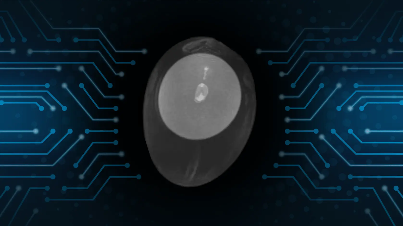
At Orbem, we’ve developed an AI-powered egg sorting system for in-ovo sexing, leveraging Deep Learning (DL) with an ever growing data set of hundreds of thousands of 3D MRI images of eggs. Our Genus Focus system is trained to automatically recognize patterns in these images and classify them as male or female.
The process involves showing MRI images of eggs to the AI, labeled as “male” or “female.” From these examples, the AI learns to identify subtle patterns in internal egg structures that a human eye might miss. This training leads to a highly accurate system capable of making “intelligent” in-ovo sexing decisions.
The great power of such AI systems is that they keep learning as they see more diverse data. Our scientists carefully design our datasets to ensure the system can accurately predict across a full spectrum of biological variation, such as eggs with differing incubation durations, breeds, and providers. As we scan more and more eggs, our solution becomes more and more accurate, continuously improving and learning from its past mistakes.
Another strength of AI systems is their versatility. As we expand our dataset with MRI scans of other objects, such as fruit, the system can learn to classify them as well for any purpose that fits our customers.
AI for Faster MRI
AI plays a crucial role in accelerating MRI processes and reducing costs, not just in making informed decisions.
A significant hurdle in MRI is the extended time required to scan a single object to obtain high-resolution 3D images. However, industrial conditions demand rapid scanning, aiming for throughputs of thousands or even tens of thousands of objects per hour.
MRI scanning can be made faster by reducing the quality of scanned images, such as lowering their resolution. Another acceleration approach is to scan a smaller part of the image: if we go back to the concept of slices in a 3D image (be it of an apple, an egg, or an entirely different biological sample), we can collect only some of those slices and not the full image, thus reducing scan time, but also losing relevant information about the object. Lower-quality or incomplete images, albeit fast to scan, compromise the accuracy of classification decisions.


We faced this challenge at Orbem too. As we pushed the throughput of MRI scanning to higher values, we started seeing a drop in classification accuracy. This is where AI shines again. We invented an innovative solution in which we first perform a very fast, low-resolution scan, and use DL to find the region of interest in the egg. Then we scan only a few slices in high resolution in that region of interest and are able to classify the sample.
As a result, our DL models not only classify the sample, but also inform MRI scanners of where to look. We achieved record speed without any loss of accuracy. We are also exploring how DL can be applied to fast acquired, low-quality images and restore their resolution and other details. This approach makes sure that we maintain high throughput and preserve high accuracy of the decisions that the AI system makes.
The Orbem Advantage
At Orbem, we’re dedicated to solving problems by looking deeper. We believe that many of the world’s most pressing challenges in food and health stem from making decisions based on guesswork and external appearances. Our groundbreaking AI-powered MRI technology changes that by revealing what’s on the inside.
We provide non-invasive, industrial-scale insights that turn the invisible into actionable knowledge – whether it’s determining the sex of an embryo within an egg or assessing the quality of an avocado without cutting it open. By unlocking this previously inaccessible information, we empower food producers, researchers, and the health sector to reduce waste, improve quality, and build a more sustainable and transparent future.
Orbem integrates the power of AI algorithms into MRI systems, bringing intelligence to imaging. AI-powered imaging already enables our customers in the poultry industry to see what is hidden behind the egg shell, and make informed decisions on which eggs to incubate. This safe and automated technology holds great scaling promise: it can be extended to many other food types and even to healthcare. The potential of Orbem’s unique technology scales as we continue to make MRI even faster and more affordable, and our AI algorithms even more accurate.
About the author
Ksenia drives Orbem’s AI and Data Science strategy and growth. Before joining the company, Ksenia did research at the Max Planck Institute for Neurobiology (now Biological Intelligence) and worked on engineering challenges in the field of autonomous driving.

Ksenia Yashina
Head of AI
Other interesting articles
From Hospital to Hatchery: How We Automated MRI
A look at the robotic egg chain, intelligent data tracking, and AI-driven sorting in the hatchery.

This Is the Impact of In-Ovo-Sexing with the Genus Focus
100 million eggs have been scanned with the contactless AI-powered technology.
We Revolutionize Poultry
How Orbem reveals what is hidden behind the eggshell.
Discover other milestones
Want to write about us?
We would love to hear from you!

Barbara Jilek
PR & Content Marketing Manager